Call Us: 011 475 7654


Our hearts have four valves which allow a one-way flow of blood. There are two on the right side of the heart (the tricuspid and pulmonary valves) and two on the left side of the heart (the mitral and aortic valves).
The tricuspid valve allows blood to pass from the right atrium to the right ventricle and the pulmonary valve which opens to allow the right ventricle to pump blood to the lungs. Oxygenated blood from the lungs then enters the left heart via the left atrium from the pulmonary veins. The mitral valve allows flow from the left atrium to the left ventricle, and the aortic valve then allows blood to be pumped into the systemic circulation from the left ventricle.
Valves can become damaged from a variety of diseases, including rheumatic heart disease, congenital heart disease, infective endocarditis as well as old age degeneration. Additionally, they can leak and become very narrow. This poor functioning of the valves can lead to leg swelling, shortness of breath, palpitations, dizziness, collapse and fatigue.
At Westrand Cardiovascular Centre, we specialise in valvular assessments, particularly in complex or multiple valve problems. We do a 3D evaluation with TOE and cardiac catheterisation to fully evaluate the problem in order to provide a treatment plan.
Cardiac catheterisation is a multipurpose procedure that allows us to diagnose and treat various heart and blood vessel issues. It's performed using a catheter, or long tube, that enters a blood vessel and is guided to the heart or area of concern. During the procedure, we can apply different tests and perform treatments.
With cardiac catheterisation, we can also effectively:
Specific diseases this procedure diagnoses include:
For this procedure, you will be entirely awake but have the area numbed so as not to feel any discomfort.
We start by inserting a plastic sheath or tube into the blood vessel (the vein that leads directly to your right heart and the artery that leads to your left heart). We may access these vessels in the wrist, groin and sometimes the neck, and use continuous X-ray monitoring to track the path of the X-ray.
When checking for a blockage, we inject contrast dye to see the flow through the blood vessels. This alerts us to any narrowing or obstructions in the blood vessel that could be causing pain or complications.
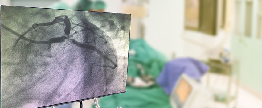
A coronary angiogram is one of the most common procedures for checking blockages or narrowed blood vessels. It uses X-ray imaging and contrast dye to show how blood pumps through your heart and body.
When arteries and blood vessels become blocked, it can lead to improper blood flow, chest pain and, ultimately, a heart attack. So it's essential to find the cause of the issue as soon as possible. A coronary angiogram is often used to diagnose blockages and determine which treatment method will be best.
We perform coronary angiograms for the following reasons:
During the procedure, we make a small incision in a central artery that connects to the heart and insert a small catheter. Then, using an X-ray monitor, we gently feed the catheter into the artery and inject contrast dye. The dye shows up on the X-ray, allowing us to see if there are any obstructions.
Book an appointment